Sunday, November 26, 2017

Hemodynamic Response to Exercise

If step one of evolutionary hemodynamic planning was maintaining blood pressure well enough to get out of bed, step two was to get us out of the house and walking. In other words, our homeostatic mechanisms have to be able to maintain adequate perfusion pressure during activity as well as during rest. The mechanisms of this process involve multiple processes which include the activity of muscle receptors, cardiac receptors, and baroreceptors, as well as feed-forward central command that originates in the higher regions of the brain.

Part 1: Baroreceptor Response
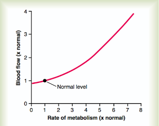
Figure 1: The local rate of metabolism can have a profound
effect on local arterial flow do the a local decrease in vascular
resistance. From Guyton and Hall (2016)
To a certain extent, the autonomic response to exercise can be thought of as an extension of the same baroreceptor response that we discussed in reference to orthostasis. In the orthostatic setting a positional drop in stroke volume resulted in decreased arterial blood pressures which stimulated the baroreceptor response. In the exercise setting, the initial drop in blood pressure is related to an exercise-induced drop in vascular resistance related to metabolic byproducts.

During muscle activity, which may range from walking to the refrigerator to running a marathon, the muscles being activated can increase their metabolic activity by 50-fold or more. In these settings of increased metabolic demand, vasodilatory metabolites are formed which include protons, potassium, phosphate, AMP, and adenosine. These substance act locally on the vascular smooth muscle which results in vasodilation. 

The vasodilation is certainly intended to increase oxygen delivery to the muscle to meet metabolic demand, but it also has the effect of reducing systemic vascular resistance (SVR). Because the mean arterial pressure (MAP) is equal to the cardiac output (CO) multiplied by the vascular resistance, this local vasodilation results in a decrease in MAP. This decrease in MAP then stimulates the baroreceptor reflex which results in modulation of sympathetic and parasympathetic tone to maintain target perfusion pressures.

MAP = CO * SVR

The increased sympathetic tone carried through local sympathetic nerves result in peripheral vasoconstriction. The intention of this reflex is to increase SVR to maintain perfusing pressure in the exercising muscle. However, the mechanism of increase vascular resistance may well backfire if the sympathetic nerves also increased vascular tone at the arterioles perfusing the metabolically active tissue. For this reason, local metabolic vasodilation takes precedence over the autonomic tone and the arterial supply to the metabolically active tissue remains vasodilated. The net result of this balance between metabolic and autonomic hemodynamic homeostasis is that the exercising muscle is perfused at the expense of other non-metabolically active tissues and that the MAP remains elevated for supply of vital organs such as the heart and brain.  

Part 2: The exercise-pressor response (EPR)

Figure 2: Metaboreceptors and chemoreceptors in the muscle
send afferent signals through group III/IV fibers that synapse
on the NTS to decrease parasympathetic in order to increase
heart rate and blood pressure. This inhibitory synapse also
results in blocking baroreceptor signals and increase the set
point of the MAP for the baroreceptor reflex.
Although the baroreceptor response is able to modulate autonomic tone through detection of vasodilation related to metabolic byproducts, this is hardly the ideal mechanism to maintain constant perfusion. After all, it requires a build-up of metabolic products, which only occurs after intense metabolic activity. The exercise-pressor response is a system of visceral afferents which are able to detect metabolic activity and adjust hemodynamics before the actual metabolic byproducts are produced that will result in the intervention of the baroreceptor reflex.

The sensors in place are not completely understood, but seem to consist of diverse metabolic and mechanoreceptors generally termed "ergoreceptors". Signal from these sensory neurons are carried to the lumbar dorsal horn of the spinal cord by thinly myelinated (group III) and unmyelinated fibers (group IV "C-fibers"). This pathway terminates on the NTS similar to the baroreceptor fibers, but exerts and inhibitory, rather than excitatory role. The net result of this is to inhibit parasympathetic tone and amplify sympathetic tone. Along with this, the effect of inhibiting the excitatory inputs to the NTS results in forcing the baroreceptor reflex to operate at a higher MAP.

Figure 3: Inputs from the EPR synapse on the NTS where inhibitory interneurons exert an inhibitory effect on the parasympathetic tone and by negating the effects of parallel excitatory input from the baroafferents, result in an increase in MAP set point for the baroreceptor response. This figure also demonstrates the same effect of central command extending through the PVN, which also results in an increase of the MAP set point. From Michellini et al. (2015) 
Part 3: The role of Central Command

Continuing with our earlier line of thought, while EPR is superior to the baroreceptor response to avoid transient hypoperfusion during early exercise, it still does require increased metabolic activity to function. The ideal homeostatic mechanism for exercise would be one that initiated tachycardia and hypertension prior to any increased metabolic activity, so that the appropriate hemodynamic reserve would already be in place once exercise had begun. This mechanism is called central command.

Because central command takes its cue from cortical centers of higher-level processing which we do not understand very well, the physiology of central command is somewhat murky. At a basic level, central command is a direct inhibitory signal to the NTS which comes proximally from the paraventricular nucleus (PVN), but reflects the integration of multiple higher-level processes.

The net result is that during moderate exercise, higher level processing causes an increase in the sympathetic/parasympathetic ratio in order to prepare for exercise. Once exercise has begun, central command and EPR adjust the baroreceptor reflex to operate at higher mean pressures. At these higher pressures, second-to-second hemodynamic fluctuations are managed by the baroreceptor reflex to keep the MAP at a constant appropriate level relative to the degree of central activation and peripheral exercise intensity.


References: 

Guyton and Hall Textbook of Medical Physiology (2016)
Michellini et al 2015


Bainbridge Reflex and Renal Sympathetic Tone

Just as moment-to-moment blood pressure control is maintained through baroreceptor-mediated cardiovascular sympathetic tone, long-term blood pressure homeostasis is controlled through renal extension of the same process. It is one of the most fascinating aspects of the neurohormonal system that even though sympathetic tone for the entire body is processed in the same brain centers, sympathetic output can be customized to each organ for precise control. 

If each organ can have a customized sympathetic output, it makes sense that each should also have a customized afferent input. Whereas the cardiovascular autonomic tone relies primarily on the baroreceptor reflex, the renal autonomic tone relies on a mechanoreceptor reflex located at the cavoatrial and pulmonary-atrial junctions.

Figure 1: Illustration of the antagonistic relationship between the baroreceptor
and Bainbridge reflexes.
The presence of mechanoreceptors in the atria has been known for some time. Early studies revealed that inflation of a balloon in the cavoatrial junction was able to decrease thirst in instrumented animals, presumably by communicating an elevated CVP to hypothalamic centers. Further studies revealed that these mechanoreceptors sent processes to the NTS, CVLM, and paraventricular nucleus, similar to the baroreceptor afferents. Unlike the baroreceptor reflex, however, which detects increased arterial pressures and stimulates a decrease in heart rate, the cavoatrial reflex detects increased central venous pressures and stimulates an increase in heart rate (termed the Bainbridge reflex). The overall purpose appears to be to clear right-sided venous congestion by increasing cardiac output.

Figure 2: Cardiac distension results in release of ANP and BNP
along with activation of the Bainbridge reflex and renal 
sympathetic inhibition. The result of these combined 
neurohormonal reflexes is inhibition in the renin-angiotensin-
aldosterone-ADH system and appropriate sodium and volume 
wasting
The relationship between the bainbridge and baroreceptor reflexes is quite complex and antagonistic. Infusion of IV saline at a high rate initially results in increased CVP and activation of the cavoatrial receptors. This leads to parasympathetic inhibition and reflex tachycardia. Once this increased venous volume reaches the left ventricle, however, it results in increased stroke volume through the Frank-Starling relationship and enhances cardiac output. The resulting increase in arterial pressures activates the baroreceptors in the carotid bodies and aortic arch, which result in parasympathetic stimulation and reflex bradycardia. Along with the baroreceptor reflex the bainbridge reflex is part of the determinants of resting heart rate in humans, and loss of either reflex results in relative tachycardia or bradycardia, respectively.

Even more interestingly, however, activation of these atrial receptors results in a profound suppression of sympathetic tone to the kidneys. Because sympathetic renal tone results in activation of the renin-angiotensin-aldosterone-ADH system, which all tend towards sodium and fluid retention, inhibition of sympathetic tone through the Bainbridge reflex results in fluid and sodium wasting and volume depletion. This is also congruent with the hormonal effect of atrial/ventricular stretch, which is release of ANP/BNP which both mediate natriuresis and promote volume homeostasis.

One interesting finding related to these reflexes is that in chronic hypertensives, the bainbridge reflex seems specifically dampened. These patients have been found to have elevated renal sympathetic tone measured by serum norepinephrine, which at high levels of activation can leak from within the sympathetic synapses. This renal sympathetic tone, however, does not correlate with the patients heart rate, suggesting that cardiac sympathetic tone is relatively depressed. The end result is the relative volume overload and increased SVR that characterizes essential hypertension.

In the context of chronic hypertension, the baroreceptor reflex is also relatively inactive. This is partly because at increased pressures vascular compliance is low and further blood pressure changes do not result in sufficient arterial stretch for detection. Along with this mechanism, chronically activated baroreceptors do undergo receptor desensitization, which has the net effect of allowing the baroreceptor to reset its basal MAP point to higher pressures for the purpose of maximizing dynamic range. Lastly, hormonal regulation of hypertension results in elevated angiotensin levels, which exert a central suppressive effect on central mediators. 

Because of the complex physiology of excessive renal sympathetic tone and resulting baroreceptor desensitization in chronic uncontrolled hypertension, a large multi-institute study (SYMPLICITY HTN-3) was performed which investigated the effect of selective renal denervation on chronic resistant hypertension in optimally medically managed patients (average of 5.1 medications). Unfortunately the trial did not demonstrate any benefit to this population, despite promising phase I and phase II studies. It's unclear why renal denervation was not more successful, although it may prove to have a more significant impact on a less heavily medicated population, or a population that is unable to tolerate medication due to side effects.

References: 

Coote, PH A role for the paraventricular nucleus of the hypothalamus in the autonomic control of heart and kidney exp physiology 90.2 pp 169-173 

Klabunde R, Cardiovascular physiology, http://www.cvphysiology.com/

Pocock SJ, Bakris G, Bhatt DL, Brar S, Fahy M, Gersh BJ. Regression to the Mean in SYMPLICITY HTN-3: Implications for Design and Reporting of Future Trials. J Am Coll Cardiol 2016;68:2016-25.

Presented by Dr. George Bakris at the European Society of Cardiology Congress, Barcelona, Spain, September 1, 2014.
Bakris GL, Townsend RR, Liu M, et al. Impact of renal denervation on 24-hour ambulatory blood pressure: results from SYMPLICITY HTN-3. J Am Coll Cardiol 2014;64:1071-8.

Bhatt DL, Kandzari DE, O’Neill WW, et al., on behalf of the SYMPLICITY HTN-3 Investigators. A controlled trial of renal denervation for resistant hypertension. N Engl J Med 2014;370:1393-1401.

Thursday, November 23, 2017

Stress transduction in the adrenal medulla

Along with the myriad of sympathetic nerves which are finely distributed throughout the body, there is also a cluster of specialized sympathetic neuroendocrine cells which comprise the adrenal medulla. Unlike the mesodermal cortex which surrounds it, the adrenal medulla is embryologically related to the same neural crest tissue that develops into the post-ganglionic sympathetic neurons, but through the course of development take on a very different character.

Figure 1: Histology of the adrenal gland. The mesodermal adrenal cortex
surrounding the medulla is divided into three functional and histologic layers.
The adrenal medulla consists of a homogenous population of adrenal chromaffin
cells, which together comprise a specialized type of sympathetic ganglion.
Part 1: Chromaffin Cells

The effector cells of the adrenal medulla are the chromaffin cells.
Unlike the typical sympathetic neurons, the chromaffin cells do not have dendritic processes and do not form synapses on target organs. Rather, when they are appropriately stimulated they release the contents of their vesicles directly into the bloodstream where they exert an endocrine, rather than paracrine effect.

Figure 2: Chromaffin cells of the adrenal medulla. These cells, were they to
undergo malignant transformation, are the precursor cells of pheochromocytoma
or sympathetic paraganglioma.
As a form of specialized sympathetic ganglion, the adrenal medulla receives its innervation directly from the cholinergic preganglionic fibers. These fibers stem from the cervical sympathetic chain at the level of T5-T8 and comprise the greater splanchnic nerve. There are also postganglionic sympathetic nerves stemming from the celiac ganglion which penetrate the adrenal gland and innervate the capillary bed, but do not interact with the chromaffin cells themselves.

Part 2: The Adrenal Synapse

The presynaptic terminal of the splanchnic nerve fiber contains both small clear vesicles (SCVs) and as well as large dense-core granules (LDCGs). Much as in other sympathetic nerves, the small clear vesicles contain acetylcholine, which is the primary neurotransmitter of the preganglionic sympathetic nerves. The large dense core granules contain a neuropeptide called PACAP (pituitary adenylate cyclase activating peptide).

As with the rest of the sympathetic nerves, the acetylcholine-containing SCVs are released proportionally to the firing frequency of the splanchnic nerve. The large dense core vesicles, on the other hand, require a profoundly increased calcium concentration for exocytosis. In order to create this large calcium influx, extremely high firing frequency is required. In this way the splanchnic nerves have both a graded response expressed in acetycholine release as well as an all-or-nothing response expressed in PACAP release. 


Figure 3: The anatomy of the splanchnic nerve and associated sympathetic fibers innervating the adrenal gland. From Netters.
Part 3: Stress Transduction

Given the physiology of the presynaptic terminal and its distinction between regular firing and high-intensity firing, it is unsurprising that the adrenal medulla has distinct ways of responding to the two firing modes in terms of its response. Low frequency firing and acetylcholine release are sufficient to produce a graded response of norepinephrine release from the chromaffin cells. Acetylcholine release does also produce a small amount of epinephrine release as well, however, due to receptor desensitization and vesicle depletion acetylcholine alone is unable to produce a robust epinephrine response, instead only producing limited transient release. 

Figure 4: Physiology of the splanchnic nerve terminal. Low firing frequency
results in release of acetylcholine from SCVs only, whereas high firing frequency
is sufficient to cause release of PACAP from LDCGs. From Smith and Eiden
(2012).
High firing frequency and PACAP release, on the other hand, produces a complex set of responses within the chromaffin cell which result in robust and sustained epinephrine secretion. This response begins with direct PACAP mediated exocytosis of epinephrine, which unlike the acetylcholine-mediated response is non-desensitizing as well as action potential independent. This response is followed by induction of multiple enzymes including tyrosine hydroxylase and PNMT which are key members of the epinephrine synthetic pathway and are required for maintained epinephrine secretion. In other words, high frequency splanchnic nerve firing represents the sympathoadrenal stress response and is required for high volume endocrine secretion of epinephrine.

Part 4: Glucocorticoids are necessary for the medullary response.

Figure 5: Activation of the adrenal cortex. From Herman et al (2016)
One final component of the adrenal medullary stress response is that, while it requires sympathetic activation in the form of high frequency splanchnic nerve firing, it also requires concurrent stimulation by the HPA axis in the form of cortisol secretion. As was discussed earlier, the adrenal cortex and adrenal medulla share very little in terms of embryology. They also differ wildly in terms of overall structure, as the adrenal cortex is a hormonal system regulated by pituitary ACTH while the adrenal medulla is part of a neural system regulated by splanchnic sympathetic tone. However, inasmuch as they comprise two arms of a physiologic stress response they have much in common and even at times have overlapping functional roles.

Much like the sympathetic response, the adrenal cortical response is initiated in the paraventricular nucleus of the hypothalamus (PVN). Also similar to the sympathetic response, activation of the CRH-secreting neurons can be activated through peripheral mechanisms, largely stimulated by the same pre-sympathetic pathways that activated the CVLM for sympathetic tone, as well as central mechanisms through the limbic system for anticipatory or psychologic stress responses. In either circumstance, activation of the PVN in this manner results in release of CRH into the hypophyseal-pituitary circulation, which results in ACTH secretion by the anterior pituitary gland. Once in systemic circulation, the ACTH activates receptors in the adrenal cortex which result in production of glucocorticoids. These glucocorticoids then drain through the corticomedullary circulation where they result in transcriptional activation of PNMT.

In patients with adrenal insufficiency the resulting low corticosteroid state prevents adequate transcription of PNMT and eliminates the serum epinephrine response to stress. In experimental models of PACAP depletion or knockout basal PNMT and epinephrine secretion still exist, but are part of a low-volume easily depleted response in the absence of an autonomic stress response.

References: 
Smith, Corey B., and Lee E. Eiden. "Is PACAP the major neurotransmitter for stress transduction at the adrenomedullary synapse?." Journal of Molecular Neuroscience 48.2 (2012): 403-412.

Wolf, Kyle, et al. "Spatial and activity‐dependent catecholamine release in rat adrenal medulla under native neuronal stimulation." Physiological reports 4.17 (2016): e12898.

Herman et al. "Regulation of the hypothalamic-pituitary-adrenocortical stress response". Compr Physiol. ; 6(2): 603–621 (2016). doi:10.1002/cphy.c150015